Welcome to our comprehensive guide on diabetic eye conditions—a crucial read for anyone with diabetes mellitus. Diabetes doesn't just affect your blood sugar; it can also have significant impacts on your vision.
The Impact of Diabetes on the Eye
Diabetes mellitus is a health condition that impairs your body's ability to use and store sugar efficiently. High levels of sugar in your blood can lead to damage throughout the body, including the delicate blood vessels in your retina. The retina, a critical nerve layer located at the back of your eye, is essential for detecting light and transmitting images to your brain, enabling you to see.
Diabetic Retinopathy: The Retinal Risk
One of the most common eye issues for those with diabetes is diabetic retinopathy. This condition arises when the high blood sugar levels in diabetes cause harm to the blood vessels within the retina. It's a serious concern because it can lead to decreased vision and, in severe cases, blindness.
The retina's health is paramount for good vision. It is a thin layer made up of nerve fibers located at the back part of your eye. Visual information is captured here first before being sent to your brain through the optic nerve. The macula, which is at the center of the retina, is responsible for your detailed vision capabilities—such as reading and writing.
Types of Diabetic Retinopathy
There are three main types of diabetic retinopathy that you should be aware of:
Nonproliferative Diabetic Retinopathy
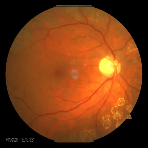
Commonly known as background retinopathy, it is an early stage of diabetic retinopathy.In this stage, tiny blood vessels within the retina leak blood or fluid. The leaking fluid causes the retina to swell or to form Deposits called exudates.
Many People with diabetes have mild NPDR, which usually does not Affect their vision.When vision is affected it is the result of Macular Edema (swelling) and/or macular ischemia (improper blood Supply).
Therefore a regular retinal check-up is necessary for all diabetics to detect the early changes.
Proliferative Diabetic Retinopathy (PDR):
This is present when abnormal new vessels (neovascularization) begin growing on the surface of the retina or optic nerve.
The main cause of PDR is widespread closure of retinal blood vessels, preventing adequate blood flow.The Retina responds by growing new blood vessels in an attempt to supply blood to the area where the original vessels closed.
Unfortunately, the new, abnormal blood vessels do not resupply the retina with normal blood flow. They are fragile and may cause bleeding in the Retina.
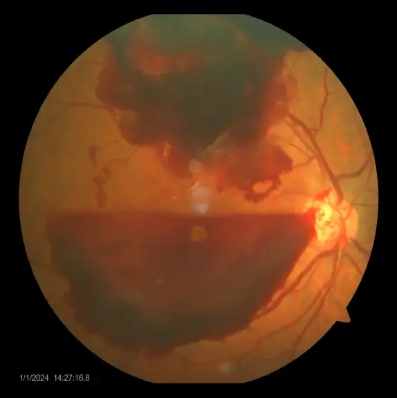
Scar Tissue that may cause wrinkling or detachment of the retina often accompanies the New Vessels. PDR may cause more severe vision loss than NPDR because it can affect both central and peripheral vision.
Proliferative diabetic retinopathy causes visual loss in the following ways:
The fragile new vessels may bleed into the vitreous, a clear, gel like substance that fills the center of the eye. If the vitreous hemorrhage is small, a Person might see only a few new dark floaters. A very large haemorrhage might block out all vision.
It may take days, months, or even years to resorb the blood, depending on the amount of blood present. If the eye does not clear the vitreous blood adequately within a reasonable time, vitrectomy Surgery may be recommended.
Vitreous hemorrhage alone does not cause permanent vision loss. When the blood clears, vision may return to its former level unless the macula is damaged.
When PDR is present, scar tissue associated with neovascularization can shrink, wrinkling and pulling the retina from its normal position. Macular wrinkling can cause visual distortion. More severe vision loss can occur if the macula or large areas of the Retina are detached.
Neovascular Glaucoma :
Occasionally, extensive retinal vessel closure will cause new, abnormal blood vessels to grow on the iris (Colored part of the Eye) and block the normal flow of fluid out of the Eye. Pressure in the Eye builds up, resulting in neovascular glaucoma, a severe eye disease that causes damage to the optic nerve.
Diabetic Maculopathy - Clinically Significant Macular Edema (CSME):
In diabetic maculopathy, fluid rich in fat and cholesterol leaks out of damaged vessels. If the fluid accumulates near the center of the retina (the macula) there will be distortion of central vision.
If too much fluid and cholesterol accumulates in the macula, it can cause permanent loss of central vision. CSME (Clinically Significant Macular Oedema) is the term given to describe water logging of the macular area .The Swelling is the most common cause of visual loss in diabetics. Most Patients with CSME need laser. Your eye care provider can see this when he/she examines your eye.
Diabetic maculopathy requires treatment if fluid is leaking into the macula. The treatment begins with identifying the leaking blood vessels on the fluorescein angiogram.
Laser treatment can be applied to seal the leaking vessels. The laser is an intense beam of light, which can be finely focused on each individual leak.
Laser is effective in stabilizing or improving vision in 75% of patients with macular edema. Despite treatment, 25% of patients continue to lose vision due to recurring leaks. Control of diabetes and blood pressure is important in reducing the chances of leaking vessels returning following treatment. The fluid often takes up to 2 to 3 months to dry up following closure of abnormal vessels.
Visual recovery is slow and gradual. If the fluid persists, the fluorescein angiogram is repeated to determine the site of the vessels still leaking and laser treatment may be repeated. The average patient needs 2-3 laser sessions per eye to control diabetic maculopathy over the course of their lifetime
How it helps you to stay strong
Make Appointment

Select Doctor
Get Consultation